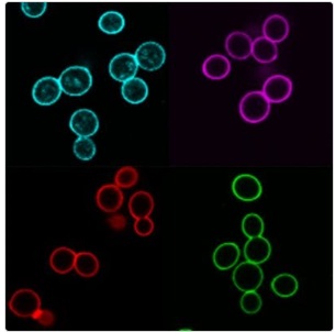
A lectin labeled with our superior fluorescent CF® dyes is an effective and widely used cell surface stain for yeast, fungi, and mammalian cells.
| Name | SKU | Size | Availability | Vendor | Price | Order | |
Concanavalin A, CF®350 Conjugate 347/448nm |
29015 | 5 x 1mg | Generally 1-2 weeks from receipt of order | Biotium | Log in for pricing | ||
Concanavalin A, CF®405S Conjugate 404/431nm |
29075 | 5 x 1mg | Generally 1-2 weeks from receipt of order | Biotium | Log in for pricing | ||
Concanavalin A, CF®405M Conjugate 408/452nm |
29074 | 5 x 1mg | Generally 1-2 weeks from receipt of order | Biotium | Log in for pricing | ||
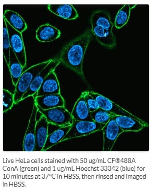
Concanavalin A, CF®488A Conjugate 490/515nm |
29016 | 5 x 1mg | Generally 1-2 weeks from receipt of order | Biotium | Log in for pricing | ||
Concanavalin A, CF®594 Conjugate 593/614nm |
29017 | 5 x 1mg | Generally 1-2 weeks from receipt of order | Biotium | Log in for pricing | ||
Concanavalin A, CF®633 Conjugate 630/650nm |
29018 | 5 x 1mg | Generally 1-2 weeks from receipt of order | Biotium | Log in for pricing | ||
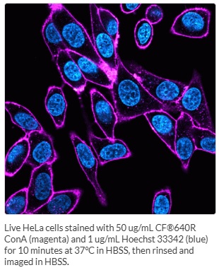
Concanavalin A, CF®640R Conjugate 642/662nm |
29019 | 5 x 1mg | Generally 1-2 weeks from receipt of order | Biotium | Log in for pricing | ||
Concanavalin A, CF®680 Conjugate 681/698nm |
29020 | 5 x 1mg | Generally 1-2 weeks from receipt of order | Biotium | Log in for pricing | ||
Concanavalin A, CF®750 Conjugate 755/777nm |
29080 | 5 x 1mg | Generally 1-2 weeks from receipt of order | Biotium | Log in for pricing | ||
Concanavalin A, CF®770 Conjugate 770/797nm |
29058 | 5 x 1mg | Generally 1-2 weeks from receipt of order | Biotium | Log in for pricing |
Photos
Features
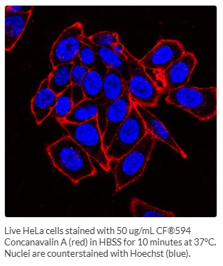
Concanavalin A (Con A) is a widely used lectin that selectively binds to the glycoproteins, a-mannopyranosyl and a-glucopyranosy. These are commonly found in the cell wall of yeast and fungi, and the cell membrane of mammalian cells and tissues.
- Stain the cell wall of yeast and fungi, and the cell membrane of mammalian cells and tissues
- Detect glycoconjugates in microscopy and flow cytometry
- Stain glycoproteins in gels
- Withstands fixation and permeabilization
- Choice of 11 CF® Dyes from UV to near-infrared
- Superior CF® Dyes are bright, photostable, and water-soluble
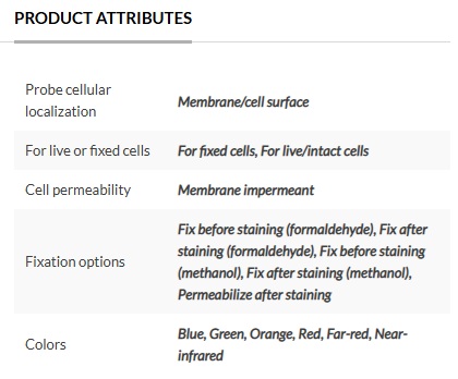
Lectins are also versatile probes for detecting glycoconjugates in microscopy and flow cytometric applications and for gel staining of glycoproteins. In neutral and alkaline solutions, Con A exists as a tetramer with a molecular weight of approximately 104 kDa. In acidic solutions (pH below 5.0), Con A exists as a dimer. Con A can be used to selectively stain the cell surface of live cells, and withstand fixation and permeabilization. When cells are fixed and permeabilized before staining, fluorescent lectins stain both cell surface and organelles in the secretory pathway.
Find the Right Stain for Your Application
Con A and other lectins are carbohydrate binding proteins that recognize specific sugar moieties on glycoproteins. The presence and distribution of these targets vary between cell types and tissues. As a result, other cell surface stains or other lectin conjugates, Wheat Germ Agglutinin (WGA) Conjugates and PNA Lectin Conjugates, may produce better surface staining and may be more appropriate for your cell type. Lectin conjugates can be used to selectively stain the cell surface of live cells, and withstand fixation and permeabilization. When cells are fixed and permeabilized before staining, fluorescent lectins stain both cell surface and organelles in the secretory pathway. Lectins may be toxic or stimulatory to live cells depending on cell type. To find the right stain for your application, see Biotium’s Membrane & Cell Surface Stains Comparison, or download Biotium’s Membrane & Surface Stains Brochure. See Biotium’s Cellular Stains Table for more information on how their dyes stain various organisms.
Superior CF® Dyes
Biotium’s next-generation CF® Dyes were designed to be highly water-soluble with advantages in brightness and photostability compared to Alexa Fluor®, DyLight®, and other fluorescent dyes. Learn more about CF® Dyes.
Note: Conjugates of blue-fluorescent dyes like CF®350, CF®405S and CF®405M are not recommended for detecting low abundance targets and may be challenging to use in tissue specimens. Blue dyes have lower fluorescence and photostability, and cells and tissue have high autofluorescence in blue wavelengths, resulting in lower signal to noise compared to other colors.
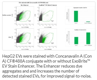
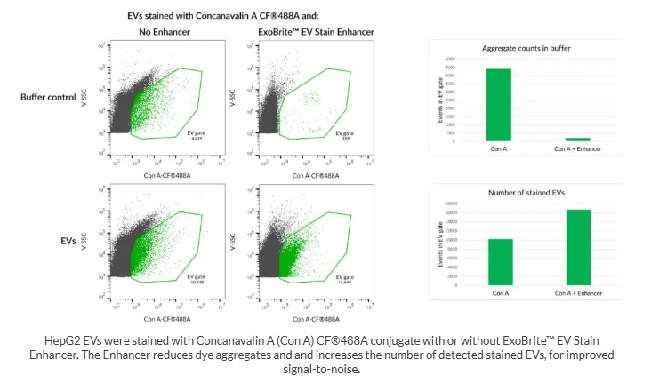
Staining EVs? Boost Your Staining with ExoBrite™ EV Stain Enhancer
The ExoBrite™ EV Stain Enhancer is a unique additive that can be added to EV stain reactions to improve the staining specificity for applications like flow cytometry. The ExoBrite™ Stain Enhancer works by reducing the aggregation of certain EV stains such as Con A and other lectins, resulting in a better signal-to-noise ratio and fewer false positives. Our testing has shown using ExoBrite™ EV Stain Enhancer with Con A staining of isolated EVs lowers aggregation and increases the number of detected EVs.
You may also like
-
ExoBrite™ EV Surface Stain Sampler Kit, Green
Kit includes each of ExoBrite™ 490/515 EV Surface Stains (CTB, WGA, and Annexin V) for assessing which stain offers the best coverage for EV samples of interest.
Log in for pricing -
ExoBrite™ Annexin EV Staining Kits
Fluorescent Annexin V conjugates that are optimized for bright and low background staining of extracellular vesicles for flow cytometry.
Log in for pricing -
CellBrite® Cytoplasmic Membrane Dyes
Dye solutions of lipophilic carbocyanine dyes DiB, Neuro-DiO, DiI, and DiD, as well as novel near-infrared lipophilic dyes for non-toxic labeling of cytoplasmic membranes.
Log in for pricing -
RedDot™2 Far-Red Nuclear Stain, 200X in DMSO
A far-red cell membrane-impermeant nuclear dye with greater nuclear specificity than Draq7™. Ideal for fixed cell nuclear counterstaining with minimal cytoplasmic RNA staining.
Log in for pricing